About David

A guy with a thing for Ice Age sloths.

Walk Like a Sloth–lesson 12: the femur

Introduction to Walk Like a Sloth: lessons in ground sloth locomotion

Getting Oriented


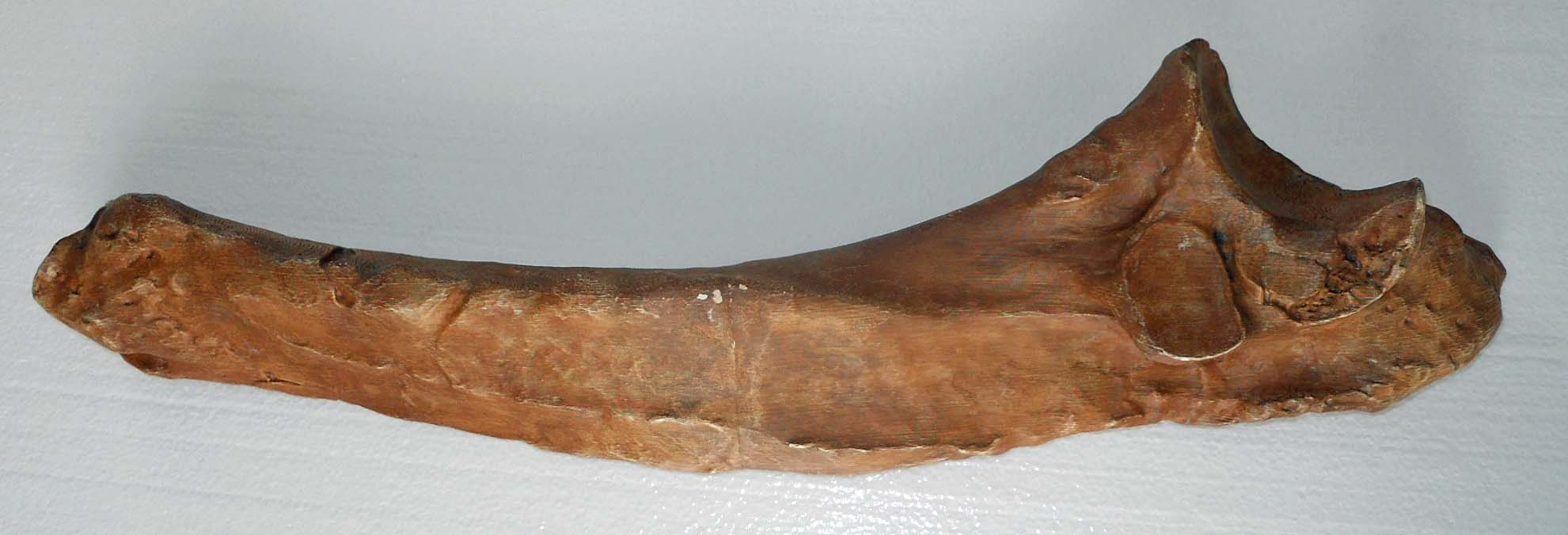
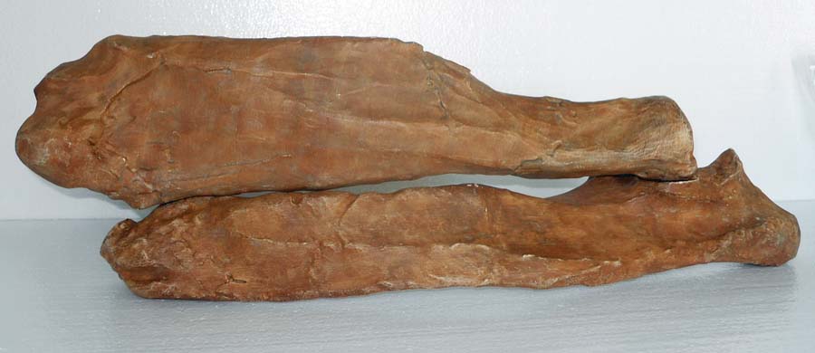
The femur or thigh bone is the largest bone in the sloth’s body and one of the most unusual bones of any animal owing to to its tremendous width and eccentric cross-section.  eccentricThe round head fits into a cup-shaped socket in the pelvis or hip called the acetabulum, while the far or distal end forms the knee joint, articulating with both the tibia or shin bone and patella or knee cap. The head of the femur points upward, about 35° below vertical, and angles forward about 45°, matching the backward-pointing hip sock.  This gives Megalonyx a unique knees-wide-apart stance.  (McDonald, 1977) The distal (down) end of the bone has a shallow wide depression in the center of one side–that’s the trochlea or patellar groove, where the patella articulates.  That’s anterior (forward) obviously, so this femur came from the sloth’s left leg.

Continue reading

Generic Levitra Online

Levitra and Levitra Orodispersible are tablets for erectile dysfunction. They are a popular alternative to other ED treatments, preferred by some men more susceptible to side effects. Levitra ODT is a type of erectile dysfunction tablet which quickly dissvoles on the tongue. The active ingredient is then absorbed into the bloodstream. They do not need to be taken with water like regular tablets do, so Levitra ODT may be preferred by those men who want a more discreet way of taking their ED medicine. Vardenafil is the working ingredient in Levitra. It functions by blocking an enzyme which can sometimes cause muscle walls to tighten and contract in those blood vessels at the entrance to the penis. – a drug designed for men with a reduced erectile function and sexual impotence. Thanks to the active ingredient vardenafil achieved by strengthening the potency and restored the health of the reproductive system. If you notice any changes in your vision while taking this drug, notify your prescriber or health care professional as soon as possible.

Walk Like a Sloth–more things to do with the scapula

magnifyingglassLook closer

The glenoid cavity is pear-shaped, wider at the bottom and narrowing at the superior end. The oval shape is generally believed to confer a greater range of motion versus the narrower sockets of quadrupeds.  (Aielo and Dean, 1990)  Note the glenoid notch midway up the ventral rim of the socket of the adult’s bone.  About half (55%) of humans have them too. (Prescher and Klümpen, 1997) Scientists disagree about their purpose.  The ventral surface of the scapula or subscapular fossa gives origin to the broad subscapularis muscle, part of the rotator cuff  quartet, which gathers as a wide tendon wrapping around the glenoid cavity to insert on the lesser tubercle of the humerus.  The notch may simply form due to the pressure of a tendon and subsequent atrophy of the bone. (ibid) Alternatively, it may provide extra surface area for anchoring the inferior glenohumeral ligament, one of ligaments surrounding the cavity, important for stabilizing the humerus head (Miles, 1997).

Continue reading

Walk Like a Sloth–lesson 10: the scapulae

Introduction to Walk Like a Sloth: lessons in ground sloth locomotion

compassGetting Oriented


The scapula or shoulder blade is a large, thin, relatively flat bone. Together with the clavicle (collar bone) and sternebrae (breast bones), the scapula is at the center of the sloth’s so-called shoulder girdle, playing an essential role in controlling movement of the upper arm as well asi_Beam_drawing_large magnifying the arm’s power.   The smoother, flatter side is the ventral or “in” side, configured to fit snugly against the ribs on the back.  The scapular spine marks the dorsal or “out” side, serving to bring  rigidity to this otherwise amazingly thin bone, like the upright part of an “I” beam.

Continue reading

Walk Like a Sloth–lesson 9: the clavicles

Introduction to Walk Like a Sloth: lessons in ground sloth locomotion

compassGetting Oriented

The clavicle or collar bone connects the scapula or shoulder blade to the sternum or breast bone. Along with the scapula and proximal end of the humerus (upper arm bone) the clavicle makes up the sloth’s so-called shoulder girdle and plays an essential role in helping control the sloth’s arm movements. The larger rounder end of the clavicle connects to the manubrium of the sternum while the smaller thinner end connects to the acromion of the scapula. Both ends are rough and pitted, not smooth as on other joints, serving to better anchor important ligaments within the shoulder girdle. These are synovial joints–in life you would find a cartilage pad between the bones and a layer of fluid to lubricate and cushion the shocks these joints must absorb.

Like your clavicle, the sloth’s collar bone is curved slightly to wrap around the front of the chest (bow side out) and attach to the scapula. Your clavicle bends forward slightly again, making an “S” shape, Megalonyx’s doesn’t, but there is great variability in shape among humans as well as sloths. (Aiello and Dean, 1990)  With the medial or “in” side versus the lateral or “out” side determined, we just need to identify the top or superior side from the inferior side or “bottom.” Look for the cone-shaped  conoid tubercle about 1/3 of the way from the thinner (acromial) end. That goes “down.”  It anchors a ligament that attaches the clavicle to the coracoid process of the scapula.  Note the anterior expansion of the clavicle at the lateral end.  That’s extra surface area for attaching part of the deltoid muscle.

sloth clavicles300

SLOTH CLAVICLES, adult (top), Toddler (bottom)

Continue reading

Walk Like a Sloth–lesson 8: teeth

Introduction to Walk Like a Sloth: lessons in ground sloth locomotion

compassGetting Oriented

tooth section

SLOTH CANINIFORM, cross section

This caniniform tooth (“in the form or shape of a canine”) is from the sloth’s lower right jaw or mandible.  The sloth had four (4) caniniforms (sometimes called “tusks”)–two (2) on the bottom and two (2) on top.  Slip the tooth out of its alveolus (socket).  The occlusal surface  (top, biting surface) is smooth and slopes toward the tongue or lingual side and away from the outside or labial side. The root is open because this tooth is ever-growing.  We can’t show you with the plastic prototype, but the tooth is composed of two layers of dentine covered with an outer layer of cementum.  The dentine layers are soft and wear away more quickly than the hard cementum layer as the sloth chews, leaving a hard raised edge for cutting.

caniniform300

SLOTH CANINIFORM, LOWER RIGHT, ADULT
view from lingual side, root on left

Continue reading

Walk Like a Sloth–lesson 7: the mandible

Introduction to Walk Like a Sloth: lessons in ground sloth locomotion

compassGetting Oriented

Only part of the mandible is in the traveling trunk–the chin and right lower jaw of the adult. The large alveolus or cavity on the superior (top) surface holds the front tooth or tusk, called the caniniform (“in the form or shape of a canine,” see tooth lesson).  On the other side of the chin there’s part of the corresponding alveolus for the left caniniform.  The narrow inward sloping shelf of bone anterior and between the front teeth is called the mandibular or predental spout.  The teeth behind the caniniforms are called molariforms (“in the form of a molar”).  A gap or diastema, separates the caniniform from the first molariform.   Notice how deep the alveoli are and how close they approach the bottom of the jaw.  These are long, deep teeth–the term for that is hypsodont–more on that in the tooth lesson too.  The small hole at the side of the chin is the mental foramen which provides an exit for the mental nerve and blood vessels that serve the front teeth, chin and lower lip. There’s another exit on the opposite side of the jaw.  The mandible has (4) teeth on each side—one (1) caniniform and three (3) molariforms.

mandible300

MEGALONYX MANDIBLE,
right anterior

Continue reading

Walk Like a Sloth–lesson 6: sternebrae and sternal ribs

Introduction to Walk Like a Sloth: lessons in ground sloth locomotion

compassGetting Oriented

SLOTH STERNUM,ventral view (Owen, 1842)

SLOTH STERNUM,ventral view (Owen, 1842)

Megalonyx has a sternum or breast bone composed of a stack of eight (8) separate bones called sternebrae  (singular is sternebra).  The sternebrae connect to the ribs through eight (8) pairs of bones called sternal ribs that curve around the front of the chest and connect to the true ribs with a thin layer of cartilage.  The triangle wide, flat, smooth back of the sternebrae is the “in” side or dorsal surface. Notice the protruding opposite side, called the ventral process, is heavily textured.  As in all Megalonyx bones, this increases the surface area for anchoring the sloth’s powerful muscles, in this case, the pectorals.

Continue reading

Walk Like a Sloth–lesson 5: the astragalus

Introduction to Walk Like a Sloth: lessons in ground sloth locomotion

compassGetting Oriented

 The astragalus (called the talus in humans) is basically the fulcrum of a lever rocking on the calcaneum or heel bone like a teeter-totter, moving the foot up and down when muscles pull on the Achilles tendon. valentine
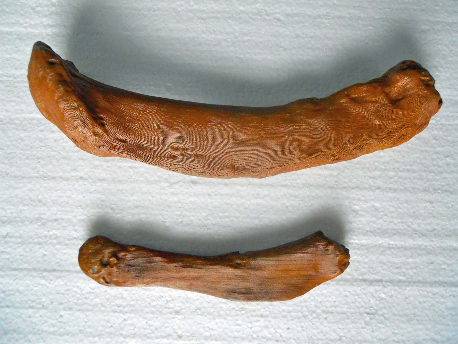
The astragalus has a convex side (the superior or “up” side) and concave (inferior or “down”) side.  With the convex side up you’ll see a heart-shaped articulating surface—orient the bone so it looks like a Valentine’s Day heart.  The smooth surface on the left side of the heart wraps around the bone and makes a right angle down to the bottom.  The fibula articulates on the left (lateral) side of the heart, extending down the outside of the astragalus, providing stability with its projecting tip. The tibia sits on the right (medial) side of the astragalus providing the main support for the leg column.  A pear-shaped cupped depression projects forward and slightly to the right pointing to the sloth’s big toe—that articulates with the navicular bone, which serves as the base for toes 1-3.  Extending laterally (to the left) from the navicular cup is the articulation for the cuboid bone, which connects to toes 4-5.  Turn the bone over.  The astragalus rocks on the calcaneum in a concave saddle formed by two articulating surfaces– a large one posterior and a smaller one anteriorly and medially (on the left side now).  Note that the joint runs diagonally across the bottom– the astragalus rides “sidesaddle” on the calcaneum. leidy calcaneumThe deep groove separating the front and back joints is called the interosseus groove or sulcus tali.  It lines up with a corresponding groove on the calcaneum forming a deep bony canal called the sinus tarsi. The rough texture of the canal serves to anchor a strong inch-wide ligament called the interosseous talocalcaneal ligament (ITCL) which ties these two critical foot bones together.

Continue reading

Walk Like a Sloth–lesson 4: the claw

Introduction to Walk Like a Sloth: lessons in ground sloth locomotion

compassGetting Oriented

The claw is from the middle toe of the left foot.  It’s the largest of all of the claws.  In life the bony core would have been covered with a toenail growing out of the pocket or sheath at the wide end. The nail probably didn’t extend far beyond the tip.  Sloth claws didn’t retract like a cat’s so a long nail would get in the way of planting the foot down flat and would have abraded quickly. Half of the nail sheath is missing from the specimen (broken off after burial) but the accident reveals the impressions left by the major blood vessels feeding the nail and penetrating into the interior through foramina (holes).  The ventral tuberosity would have supported a tough pad of tissue on which the sloths walked, lifting the tip of the claw slightly off the ground. Continue reading

Walk Like a Sloth–lesson 3: the radius

Introduction to Walk Like a Sloth: lessons in ground sloth locomotion

compassGetting Oriented

 The lower arms of mammals have two bones–the radius on the thumb side and the ulna on the little finger side. The narrow end of the radius (also called the head) is the proximal or  near end.  If it looks like a small wheel, it is! The wheel turns in the radial notch of the ulna. Note that the articulating surface on the head of the radius covers about 75° of a circle; this wheel covers about 180° on the human radius, indicating more flexibility in humans than in Megalonyx.  The large end is the wrist end; it has a small rough bump where a tendon anchors it to a corresponding spot on the end of the ulna.  The shaft of the radius is notably triangular with a prominent interosseous crest (literally “between bones”) anchoring the muscles used to turn the paws up and down. 

SLOTH RADIUS

SLOTH RADIUS, left, view from front

SLOTH FOREARM, RADIUS (top) ULNA (bottom)

SLOTH FOREARM, RADIUS (top) ULNA (bottom)

HUMAN FOREARM (left), view from back

HUMAN FOREARM (left), view from back (Clipart courtesy FCIT)

SLOTH FOREARM (LEFT), VIEW FROM FRONT  (Clipart courtesy FCIT)

HUMAN FOREARM (left), view from front (Clipart courtesy FCIT)

Continue reading

Walk Like a Sloth–lesson 2: the ulna

Introduction to Walk Like a Sloth: lessons in ground sloth locomotion

compassGetting Oriented

The lower arms of mammals have two bones–the ulna, which is located on the little-finger side of the arm, and the radius which sits on the thumb side. The narrow end of the ulna (called the head) is the distal or wrist end; the wider end with the notch is the proximal or elbow end.  The notch is called the greater sigmoid cavity or semilunar notch (literally “half-moon”), it rotates on the humerus letting you move your forearm up and down.  The projecting bone above and behind the notch is the elbow or olecranon.  A small depression below the semilunar notch and to the side is called the lesser sigmoid cavity or radial notch, where the radius articulates with the ulna.  A rough bump on the wrist end marks the anchoring point for a tendon that connects the two bones in sloths.

sloth ulna hi

SLOTH ULNA , left

ulna and radius

SLOTH RADIUS (top), SLOTH ULNA (bottom)

Continue reading