Introduction to Walk Like a Sloth: lessons in ground sloth locomotion
 Getting Oriented
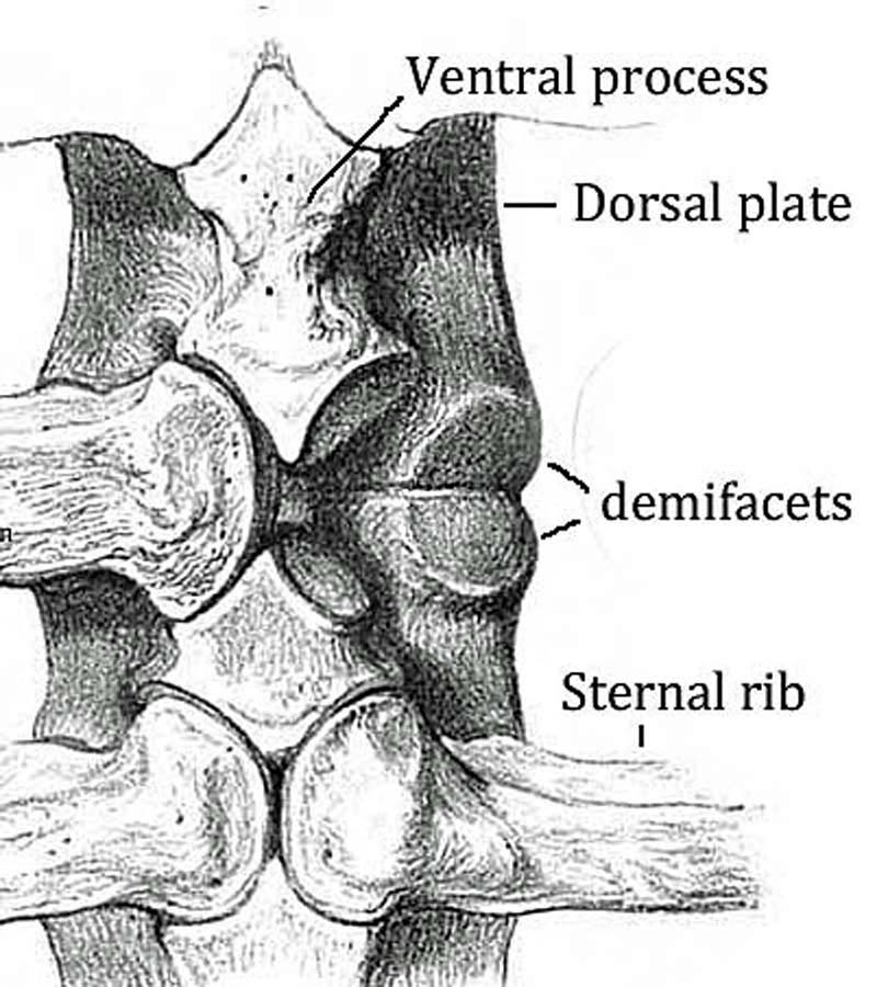
The longer, deeper-cupped joints oriented up and down are anterior (skull end), the smaller, flatter more oval joints oriented more laterally are the posterior (tail end). Now you just need to determine the back or dorsal surface from the ventral surface, to see which way the animal is facing. That’s easily done by looking for the facet inside the vertebral canal on the posterior end. That’s “down” or ventral and serves to accommodate a bony projection, the odontoid process of the next vertebra, the axis. The flattened wings projecting from the sides of the atlas are called the transverse processes. Note they are much rougher on their dorsal (top) surface, serving to anchor the muscles used to help raise and turn the head.
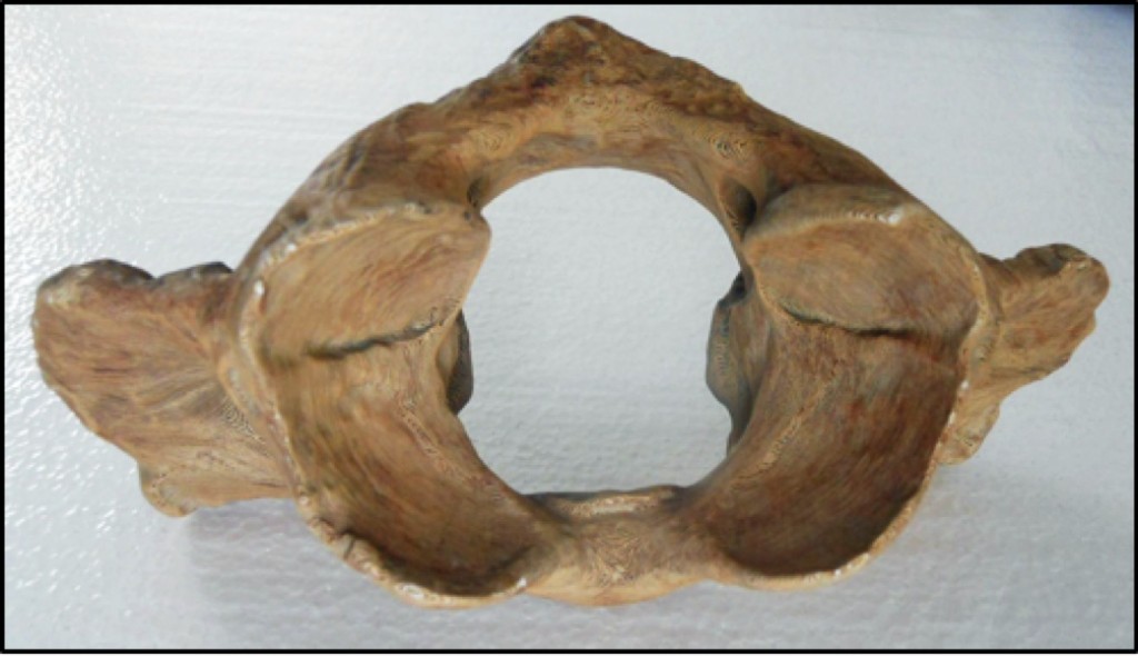
SLOTH ATLAS, view from anterior or head end
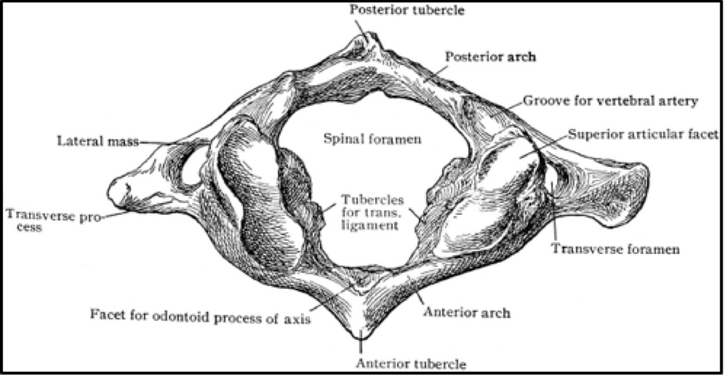
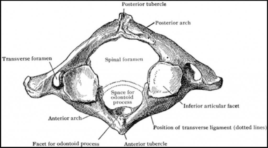
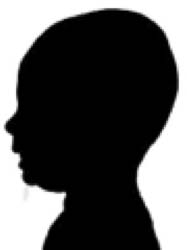
HUMAN ATLAS, view from anterior or head end (Clipart courtesy FCIT
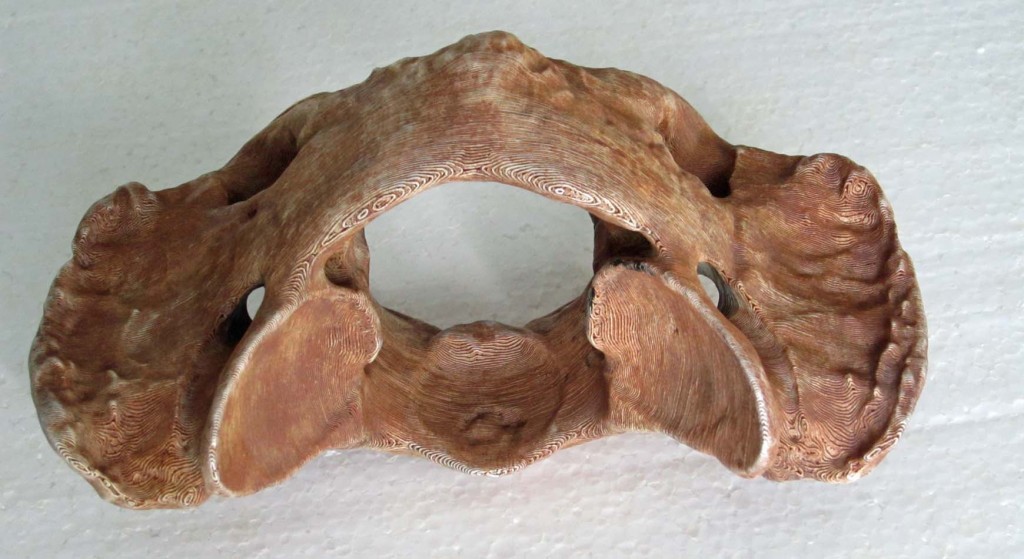
SLOTH ATLAS, view from posterior
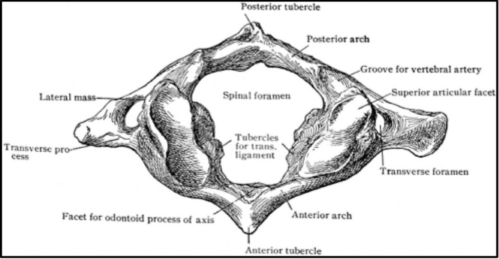
HUMAN ATLAS, view from posterior or tail end (Clipart courtesy FCIT)
Key points
#1 The atlas is the first of seven (7) cervical or neck vertebrae. It is the only vertebra that has no centrum, the bony round weight-bearing block that is the largest portion of all other vertebrae.
#2 The atlas is basically just a large ring that attaches the skull to the backbone and gives animals the ability to nod their heads up and down by rolling the skull on the front end (nodding “yes,” hence the “yes joint”). The skull turns right and left by sliding around the second neck vertebra, the axis, on the back end (shaking “no,” hence the “no joint”). Note the yes sockets are deeply concave (no lateral play in this joint) while the no sockets are relatively flat allowing sloths, like us, to turn their heads from side to side in a wide arc—there’s a much broader side-to-side motion than up and down. . . the better to watch for danger over your shoulder.
#3 There’s no room in the atlas for a centrum for it needs a large central hole to accommodate the spinal cord at its thickest point—where it exits the brain. [Compare this to the size of the spinal cord down at the back end.] The large holes along each side of the atlas, snaking over and through the transverse processes, are called the transverse foramen—these canals are found only in cervical vertebrae. They protect the vertebral arteries that transport blood to the brain. Two smaller foramina on each side allow passage of blood vessels into the nerve cord and the bone.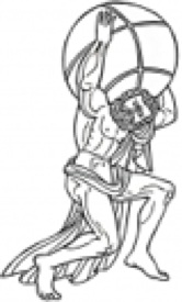
 Additional information
The atlas is named for the Greek god Atlas who was tricked by Hercules to hold up the heavens. He is commonly portrayed supporting the Earth on his shoulders.
Early observers were struck by the great length, width and thickness of the sloth’s transverse processes–evidence of great muscular forces moving the head. (Owen, 1842) Also note the roughness of the dorsal anterior surface of the atlas. The ridges anchor one of the muscles that helps raise the head. A sloth’s head, like ours, is heavy in proportion to the body so these muscles need to be large and strong. Most of the head-lifting is done by the larger nuchal muscles running from the other cervical vertebrae, collar bones, ribs and shoulder blades to the back of the skull.
The broad rough surface on the dorsal (top) side of the transverse processes serves to anchor additional muscles for raising the head as well as some used to help turn the head which attach to the axis. The muscles
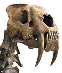
image borrowed from Wikipedia
used to lower or nod the head, the prevertebral muscles, are much smaller. Two of these attach to the underside of the transverse processes. Most mammals let gravity do most of the work of lowering the head
(which is why our head drops when we nod off) so their prevertebral muscles are relatively small.
The sabre-toothed cat is a notable exception. Their prevertebral muscles are much more developed to aid in stabbing.
Things to do
Move-your-head-like-a sloth. Use the prototype to see how the joints of the atlas work to allow sloths to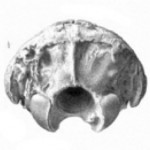 turn and move their heads “yes” and “no.” [Suggestion: use your knuckles on a couple of fingers to represent the joint with the skull, occipital condyles.]
In life, the vertebral column is wrapped in a tough elastic covering called intervertebral substance which acts like a spring holding the vertebrae together and bringing them back into alignment as you turn and move. Springing saves energy–15%, or more, so animals don’t have to use as much muscle power to hold their spines in position. (Hildebrand, 1985) But the atlas lacks the intervertebral substance—it is attached to the head and the axis with only ordinary cartilage joints and ligaments, allowing the atlas and skull to turn in any direction without resistance. Imagine how much more work it would be to turn your head if the atlas was wrapped in the tough elastic that covers the other vertebrae. [Try turning your head as though it were held by a giant rubber band (with straining sound effects of course!. Now let it spring back to the center (“boingggg.”)]
Flex Your Muscles The human atlas offers little leverage for moving the head, so most of the work is done by larger and more superficial groups of muscles attached to the torso. However, the deep small muscles serve an essential function (besides ensuring your head doesn’t fall off)—they constantly relay information to the brain about the position of your head and neck. This so called proprioceptive function may be more important than their contracting ability. (Aiello and Dean, 1990). Close your eyes. Move your head. You don’t need your eyes open to sense when your head is level. Your inner ears are helping too.
 Things to think about
Why are atlases and skulls so often missing at fossil sites? As a consequence of the lack of intervertebral “elastic” covering the atlas, the head is only loosely attached to the rest of the body. After death, the heavy skull and atlas are often the first bones to become separated from the skeleton, especially in water. In contrast, the rest of the vertebrae are often the last bones to separate or disarticulate. Finding the adult sloth’s skull at the fossil site is strong evidence the animal decayed where it died, and probably lived there–the sloths weren’t swept there by a flood, for example. Having some confidence the animals lived where they were found means the other fossils (e.g. seeds, pollen, turtle bones, etc.) found with the bones are evidence of the sloths’ habitat and wider ecosystem. What could these other fossils tells us? [Answer: nearby geography, plants, climate, season, etc.]
 Future research
Scientists are using morphometrics, a science measuring subtle differences in the shapes of bones in different species, to study the atlas and help identify different forms of locomotion. In theory, the head of a quadruped hangs from its neck requiring more robust muscular support and atlas bone structure than an animal with an upright posture (e.g. a biped, like humans) where the head is simply balanced on top. (Manfreda et al., 2006)
Conclusion
Atlases are different for every species of mammal, but all have a similar and unmistakable shape because they all serve the same basic purpose—allowing the skull to turn freely. Whether the similarity between humans and sloths is related to a common posture and mode of locomotion, or an adaptation to something else is not known and the answer awaits further research. By itself the atlas can’t tell scientists if Megalonyx is bipedal, but the similarities with the human atlas are striking.
To learn more about or to borrow the University of Iowa Museum of Natural History Geo-2-Go Discovery Trunks call or contact the museum.
References
Aiello, L. and Dean, C. 1990. Introduction to Human Evolutionary Anatomy. Academic Press Limited. San Diego, CA
Educational Technology Clearinghouse, Florida Center for Instructional Technology, College of Education, University of Southern Florida http://etc.usf.edu/clipart/
Hildebrand, M. 1985. Walking and running. In Functional Vertebrate Morphology. M. Hildebrand, D. M. Bramble, K. F. Liem and D. B. Wake (eds.) Harvard University Press, Cambridge, MA.
Manfreda, E., Mitteroecker, P., Bookstein, F. L., and Schaefer, K. 2006. Functional morphology of the first cervical vertebra in humans and nonhuman primates. The Anatomical Record Part B: New Anat.): 2898: 184-194.
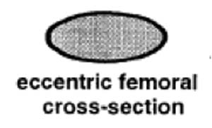 The round head fits into a cup-shaped socket in the pelvis or hip called the acetabulum, while the far or distal end forms the knee joint, articulating with both the tibia or shin bone and patella or knee cap. The head of the femur points upward, about 35° below vertical, and angles forward about 45°, matching the backward-pointing hip sock. This gives Megalonyx a unique knees-wide-apart stance. (McDonald, 1977) The distal (down) end of the bone has a shallow wide depression in the center of one side–that’s the trochlea or patellar groove, where the patella articulates. That’s anterior (forward) obviously, so this femur came from the sloth’s left leg.
 Look closer
 magnifying the arm’s power. The smoother, flatter side is the ventral or “in” side, configured to fit snugly against the ribs on the back. The scapular