Introduction to Walk Like a Sloth: lessons in ground sloth locomotion
 Getting Oriented
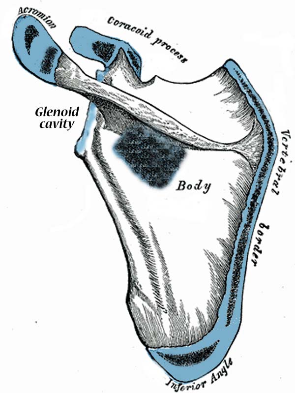
The scapula or shoulder blade is a large, thin, relatively flat bone. Together with the clavicle (collar bone) and sternebrae (breast bones), the scapula is at the center of the sloth’s so-called shoulder girdle, playing an essential role in controlling movement of the upper arm as well as magnifying the arm’s power. The smoother, flatter side is the ventral or “in” side, configured to fit snugly against the ribs on the back. The scapular spine marks the dorsal or “out” side, serving to bring rigidity to this otherwise amazingly thin bone, like the upright part of an “I” beam.
The spine arises from the vertebral border of the scapula and extends laterally as it broadens into a projection called the acromion process that bends upward and over the glenoid fossa or cavity where the head of the humerus articulates. In sloths a bridge of bone grows from the end of the acromion and meets the coracoid process projecting from the ventral surface of the scapula near the glenoid head. The combined processes and bridge are called the coracoacromial arch. 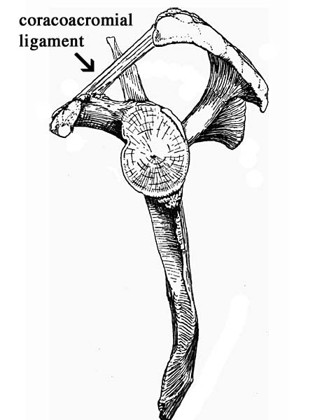 This bony bridge is unique to sloths and close relatives. In hominoids (i.e. humans and apes), and them alone, the bridge is replaced by a strong ligament. Whether bone or ligament, the bridge provides a protective covering for the head of the humerus and locks it down to prevent dislocation upward (toward the head) when the arm pulls down laterally. (Ciochon and Corruccini, 1977) No other mammals make that movement, called adduction, with force enough to need this reinforcement. In the case of sloths, the bridge also provides extra attachment area and a better angle for using their important deltoid muscles, which cover the clavicle, proximal scapula and humerus. More on that below.
In life, the glenoid cavity of the scapula points up and slightly to the side on which the scapula belongs, between one and two o’clock (20-30° from vertical). (McDonald, 1977) The cavity is much smaller than the head of the humerus, so small in fact that when ground sloths were first discovered, scientists thought the bone couldn’t possibly belong to this large animal! (Owen, 1842) A small glenoid cavity increases the range of possible arm movements, making food-gathering, for example, more efficient. But it also leaves the joint more vulnerable to injuries like strains, torn muscles and dislocations. Consequently, it is braced with an abundance of muscles and ligaments, and, as previously mentioned, a unique adaptation (the coracoacromial arch) to hold things together svenskacasinon24.se.
Also helping to identify right versus left is the coracoscapular foramen (hole) located at about two o’clock from the glenoid cavity. That’s corresponds to a notch on your scapula, called the suprascapular notch (“supra” meaning above or on top). You’ll see the foramen is also just a notch on the juvenile scapulae too. The top edge closing the foramen was still growing and hadn’t yet fused to the blade (more on that below). The notch/foramen provides a passage for the suprascapular nerve going to the muscles on the blade.
The broad flat areas or fossae (plural of fossa) that make up the blade of the scapula between the ridges serve to anchor the four short rotator cuff muscles that insert around the humerus head. The bone is almost paper thin in the center, translucent in fact, showing that stabilizing and controlling the humerus head is the priority in this instance, rather than exerting great leverage. The secondary structural thickenings on the ventral surface of the scapula divide the blade into a series of isosceles triangles any structural engineer would admire, all pointing at the glenoid fossa. The thickened ribs provides extra surface area for the rotator cuff muscles and additional stiffening against bending. The real power muscles, i.e. the ones that rotate the scapula, originate on the vertebrae and ribs and insert on the thick spine and raised edges of the scapula where they can make stronger attachments and have better leverage.

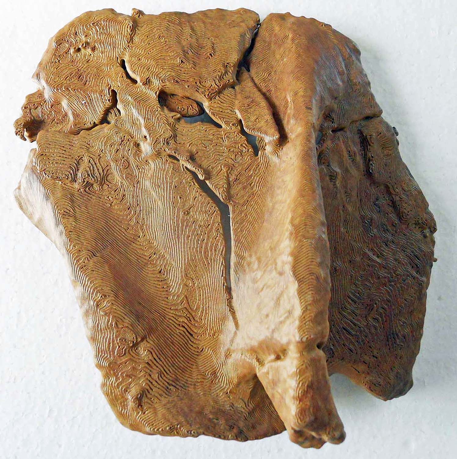
SLOTH SCAPULA, “BABY” (R),
view from dorsal side
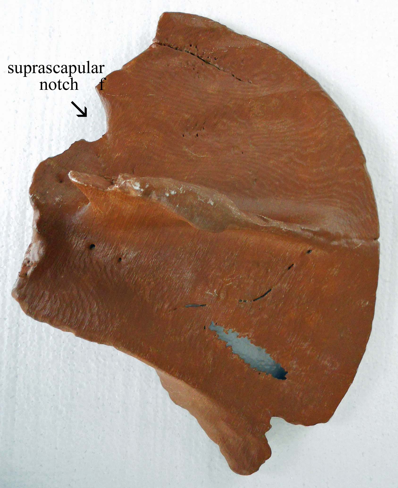
SLOTH SCAPULA, “TODDLER” (L),
view from dorsal side
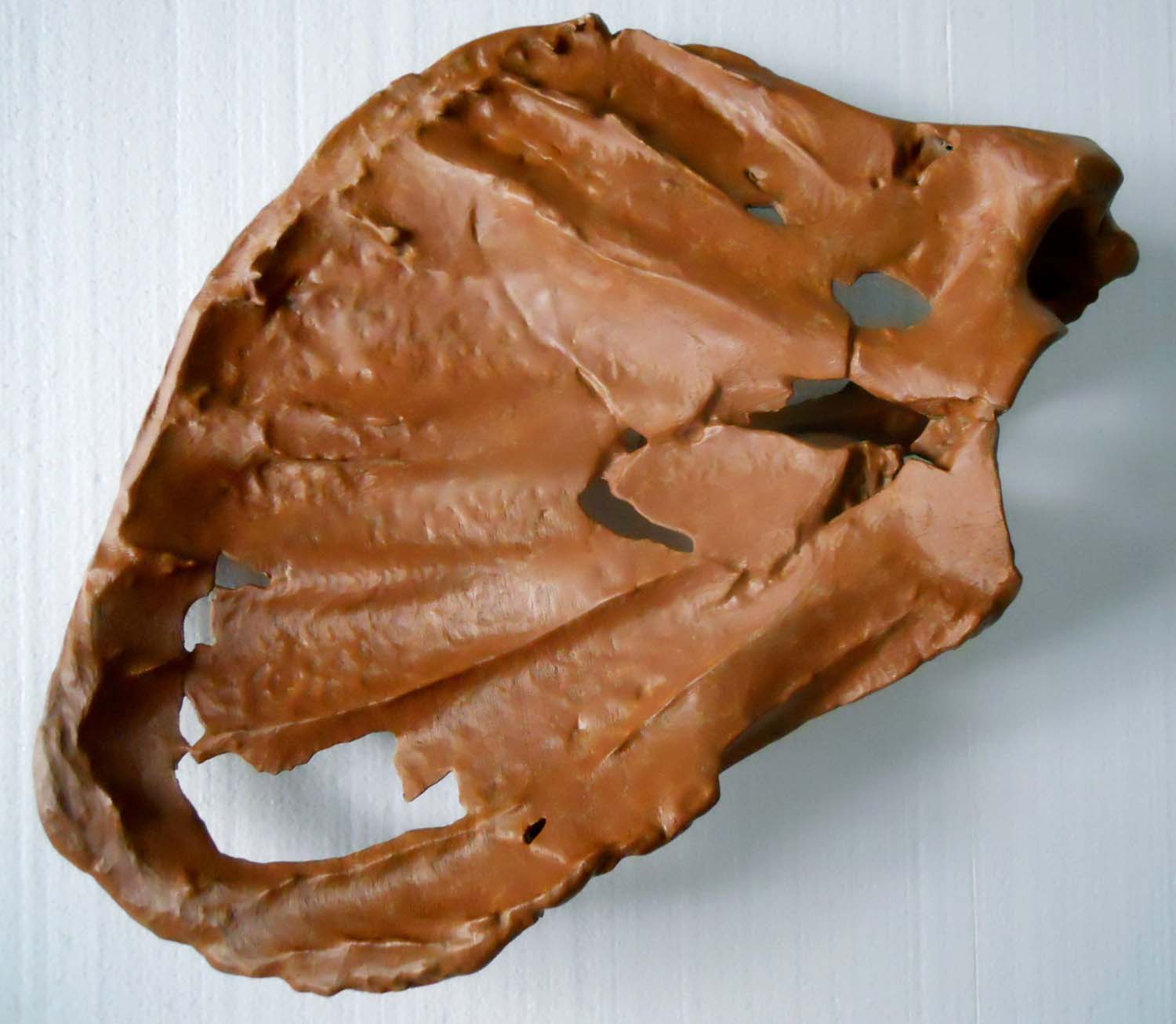
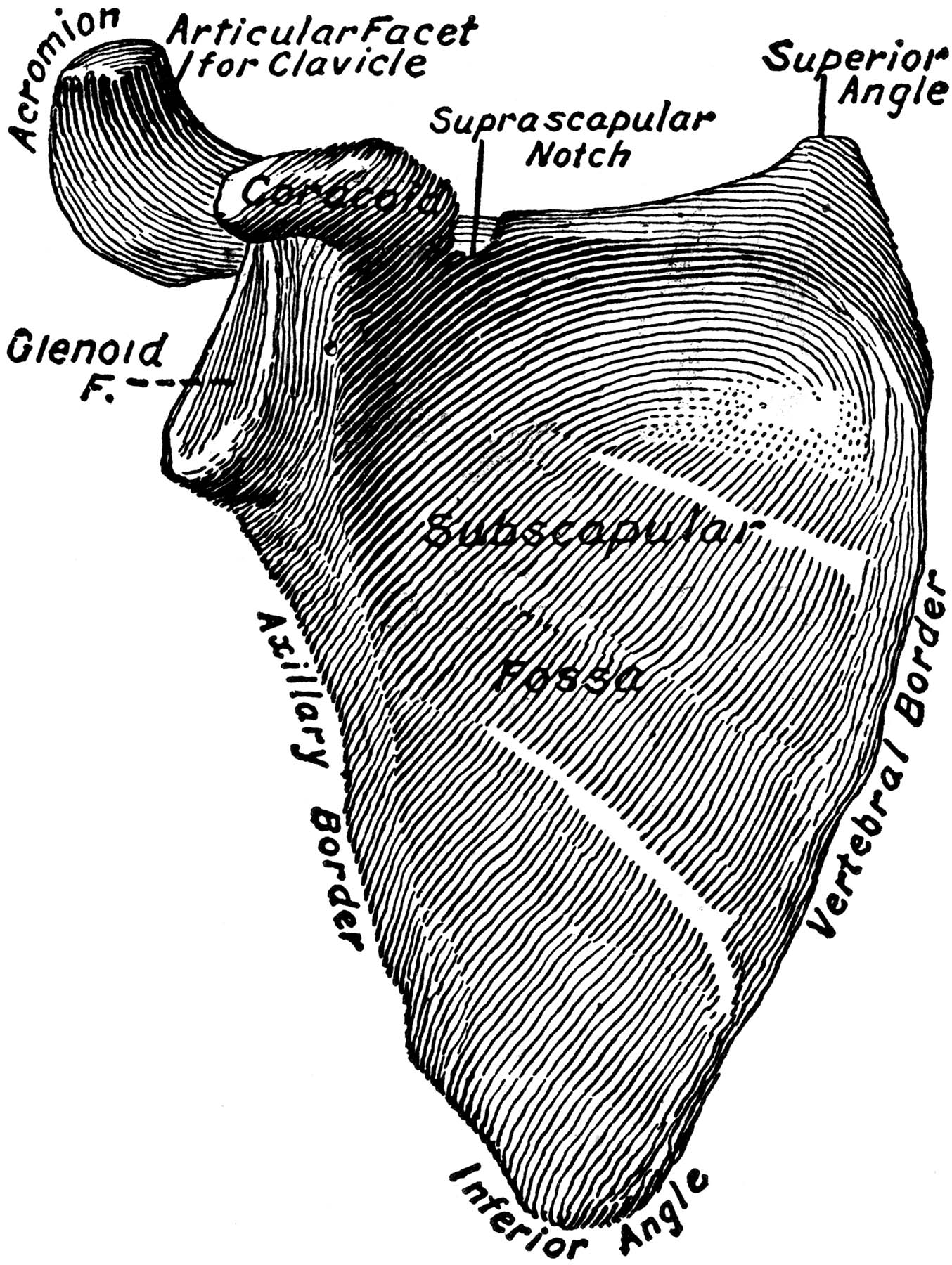
SLOTH SCAPULA, ADULT (L),
view from front
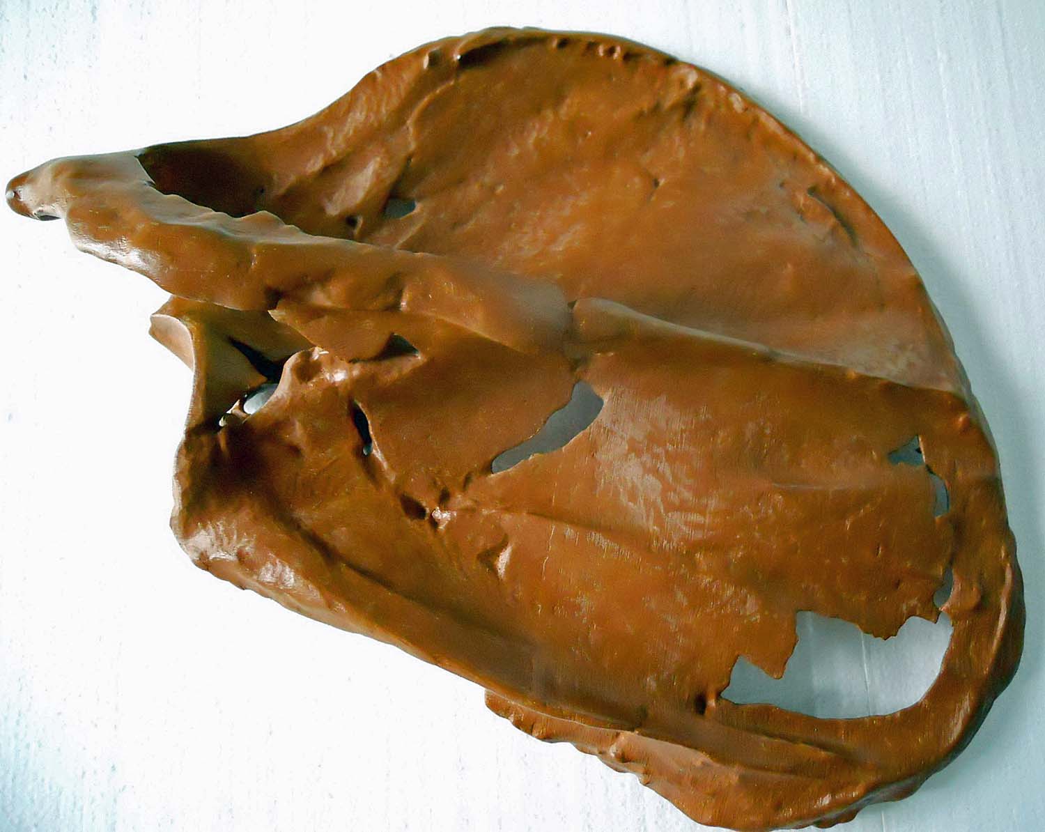
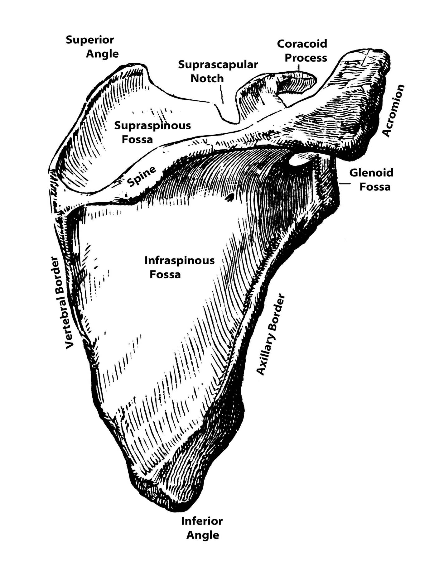
SLOTH SCAPULA, ADULT (L),
view from back
HUMAN SCAPULA (R),
view from front
(clipart courtesy FCIT)
HUMAN SCAPULA, (R),
view from back
(clipart courtesy FCIT)
 Key points
1. The only connection of the arm to the rest of the skeleton is through the scapula and clavicle, with their associated ligaments, to the sternum. The sloth’s shoulder isn’t a true floating shoulder like those of horses for example (they don’t have clavicles at all!), but like theirs (and ours too), these scapulae do float, just to a lesser degree. Shrug your shoulders. Your arms didn’t move your shoulders–they just went along for the ride. Your scapulae moved. A lot! But these are no mere corks floating in water, the scapulae are suspended in masses of powerful muscles inserting on the edges and scapula spine and rotating the bone, literally cranking it like the crank gear of a bicycle, to increase the power of of arm movements, in some cases by almost 50%. (Inman et al., 1944)
2. The scapula is a complex bone with many different forces acting on it. Seventeen (17) different muscles pull on it from different directions to perform a variety of different activities including locomotion, food gathering, load carrying, digging, defense and offense, etc. (Davis, 1964) Mammals have evolved considerable muscle redundancy in the shoulder girdle so if one muscle is strained, another can fill in. All of this makes it extremely challenging to link the shape of the scapula to specific functions, but a few general rules apply. See #3 below, for example.

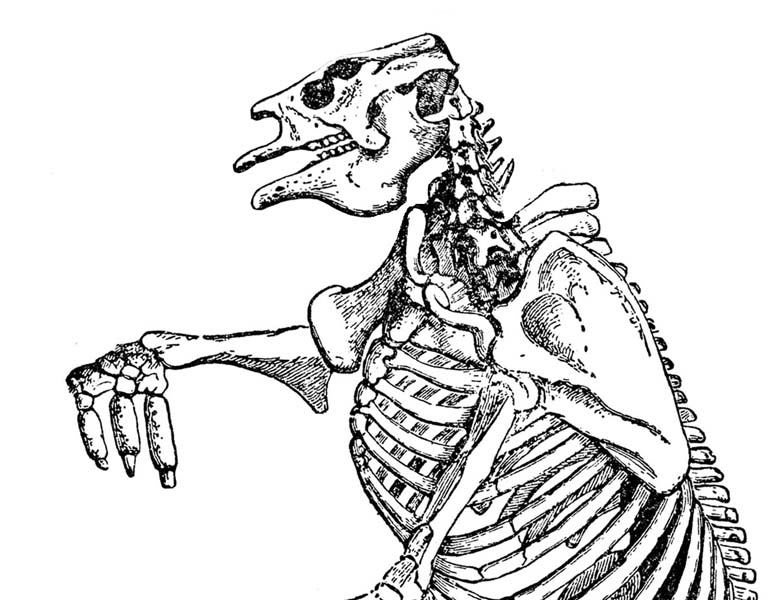
Horse skeleton.
(Clipart courtesy FCIT)
3. The scapulae of quadrupeds are generally much longer than those of bipeds; they are also positioned more laterally, that is on the sides of the animals’ rib cages. There, by moving with the limb, indeed by helping to power it, a quadruped’s scapula contributes to a longer, stronger stride. The lengthened scapula blade, and especially the edges of the infraspinous fossa, provide the needed surface area for attaching the rotation muscles. The scapulae of bipeds and arboreal (tree-living) primates are shorter and wider and shifted medially to the back, closer to the spinal column. There they can accommodate a greater range of arm motions, especially laterally (to the side), and the mass of bones and muscles is closer to the center of gravity–a safer, more stable position for animals climbing around in trees or walking upright. The supraspinous fossae are expanded to manage the more complex arm movements which include sideways as well as forward. In bipeds, however, scapulae are more than just heavy masses of muscle and bone powering the arm and threatening to tip one over at any time. Connected firmly to the trunk through the clavicles, a biped’s shoulders swing with every step and by shifting weight help to maintain balance. (see the clavicle lesson)
4. Finding three (3) pairs of scapulae told us we had three (3) sloths at the Tarkio Valley site. Five (5) of these thin and fragile bones are almost complete, except for the juvenile epiphyses (edges). How these fragile bones survived is one of the many mysteries we have about the site.
 Additional information
Scapula is derived from the Greek 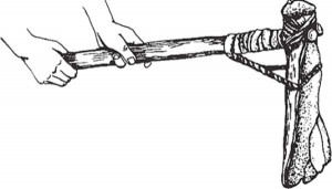 “skapto,” or “I dig,” a reference to the use of the shoulder blades of large mammals as spades and hoes before the invention of metal tools. Fossa is Latin for “ditch” or “trench.” In bones it refers to depressions or hollows where other bones articulate or muscles attach.
We generally credit the ball-and-socket of our shoulders for the tremendous flexibility we enjoy at this joint, but it’s really four (4) joints all working together. One between the sternum and clavicle (called the sternoclavicular or SC joint); a second between the clavicle and acromion (the acromioclavicular or AC joint); a third between the glenoid fossa of the scapula and the humerus (the glenohumeral joint), and finally; a fourth between the scapula and the back/chest wall (the scapulothoracic joint). Basically we can move our arms 180° in any direction, but only about 2/3 of that movement comes by virtue of the ball-and-socket/glenohumeral, the remainder comes from the other joints of the girdle. (Inman et al., 1944)
Human scapula growth centers. Image courtesy ELC.
A problem with comparing the juvenile scapulae to the adult’s is that the former include just the main body or diaphysis of the bone. The missing pieces and edges (epiphyses) (e.g. the upper glenoid cavity, coracoid, etc.) didn’t break off–they simply hadn’t fused to the main body yet and thus were easily separated after death. Vertebrates have adapted to the complex demands placed on their bones by extending their growth in important areas. Scapulae have evolved one of the most complex growth patterns of all. Human scapulae have at least eight (8) separate growth centers, and those pieces don’t completely fuse to each other until we are adults and full-grown. Growth begins in the scapular neck and coracoid. Other centers of ossification develop later elsewhere in the coracoid, plus the acromion, the glenoid cavity, the inferior angle of the body and the medial (vertebral) border. (Bass, 1971) [Scientists don’t name the landmarks on a bone just to make life difficult for anatomy students—they are in fact often separate bones while the animal is growing.

SLOTH SCAPULA, “BABY” (R),
CT scan, view from front
and key points of muscle and tendon attachment.] Continuing to grow enables bones to adapt to the changing forces around them. The epiphyses of decaying animals detach and can be easily washed away or get overlooked by untrained diggers. We were fortunate to recover one scapula epiphysis–the inferior (lower) angle on the “Toddler’s” left side.
In some sloth relatives (e.g. armadillos and anteaters) and other mammals (e.g. gophers, bears, etc.) the scapula blade is expanded posteriorly beyond the second (lower) spine into another wide fossa. That provides extra attachment area for the muscles that pull down the scapula and rotate it laterally. That’s an adaptation for digging, or climbing in the case of bears. (Davis, 1949) The absence of the expanded fossa in Megalonyx is a good clue that digging and climbing were not a major activities in their lives. Their short elbows are another indicator (see ulna lesson).
 Things to do
Math Fun: How tall were the sloths? The scapulae are the only bones we found from all three (3) sloths and the only confirmed bones we found belonging to the “Baby,” so they are the specimens we must use if we want to try to calculate the sloths’ relative sizes. Scapulae aren’t normally used for estimating mass or stature–they aren’t directly involved in height, of course. Long bones, leg bones especially, are generally preferred, but they aren’t always recovered or complete, while parts of the scapula, especially the glenoid cavity, can be very durable. Italian researchers developed formulas for estimating stature from bone fragments including the scapula, when long bones are not available. (Campobasso et al., 1998) It’s important when using any stature formulas to limit their use to relevant individuals, which in this case is contemporary adult Italians, hopefully of the same sex, age and ancestry and species. But just for fun, let’s pretend our sloths grew up in sunny Italy 100 years ago and try a Campobasso equation. Use the graphic below, adopted from Burke (2008), showing the standard (human) scapula anatomical landmarks, 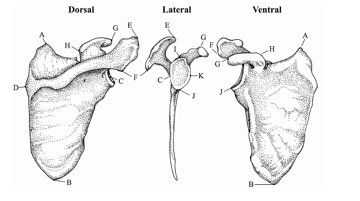 and try a measurement they found was useful for both adult males and females: the maximum width of the glenoid fossa (C-K in the diagram). Try the adult first. Compare your answer with the one you got from the arm bones and clavicle :
Height (in inches) = 56.3 in. + 8.52 X glenoid fossa width (in inches) ± 8 in.
[The glenoid fossa of the adult scapula is just over 2 1/2 inches at its widest point, so we can be 95% confident that our Italian sloth would have stood a 6 ft. 6 in. tall ± 8 inches. The arm bones and clavicle estimates suggested a stature closer to 9 feet.](see radius lesson, ulna lesson, clavicle lesson)
There’s a wider margin or error starting with this small bone, but why is there such a large difference between estimates? Which one is right? [Sloths have different proportions, and recall that we suggested before that evolution had “stretched” the forearms of Megalonyx for reaching. Maybe they weren’t as tall as the arm-numbers indicated. It’s also possible humans have evolved unusually wide glenoid fossae (Churchill and Trinkaus, 1990) and that our sloth fossa just look small in comparison. There’s also data (see more things to do with the scapula) showing glenoid fossa width is highly variable and it isn’t even highly correlated with the length of the fossa in Megalonyx. (McDonald, 1977)]
Try the stature formula on the juveniles. Calculate the heights of the “Toddler” and “Baby” sloths from the glenoid and compare your answers to the stature you calculated using the juvenile clavicle (see clavicle lesson). Can we be confident now which juvenile the clavicle came from? [No. With fossae just under 2 inches and 1.5 inches for the “Toddler” and “Baby,” the formula gives us height estimates of 6′-1″ and 5′-8″ respectively. We estimated 5′-9″ before, which is interesting, but given the margin of error, the clavicle could belong to either juvenile.] Odd, don’t you think, that the numbers came in so close together? Clearly the “Baby” is more than just 7% smaller than the “Toddler.” The width of the glenoid fossa isn’t a good predictor of stature in Megalonyx, at least not in juveniles. Compare the the three scapulae–can you see some dimensions or landmarks that might be a better height predictor? (See more things to do with the scapula for more Math Fun and to try other Campobasso formulas.)
Flex Your Muscles 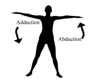 Find your collar bone and follow it out to the end of your shoulder–that bone is your acromion. Now lift your arm sideways like you were doing a “jumping jack.” That’s called abduction. Can you feel the mass of muscles at the top of your shoulder working? That’s the deltoid muscle that makes up most of the mass on top of your shoulder lifting your arm. Part of the deltoid is is anchored on the clavicle, other parts are anchored on the acromion and scapula blade (see clavicle lesson), all attaching to the lateral (out) side of the upper arm bone. Sloths used their deltoids to reach overhead to grasp tree branches. Mimic that movement.
 Pump your fist like a sloth Feel the muscle on the bottom of your arm work as you jerk it down it. That’s your triceps brachii muscle in action, or simply “triceps” to its friends. The sloth used it to pull branches off trees. The triceps has three heads or sites where it originates: the scapula–on a knob or tubercle below the glenoid fossa, and two places along the humerus, before coming together in a strong tendon that inserts on the olecranon process of the ulna (elbow). Another tubercle above the glenoid fossa anchors one head of the sloth’s (and your) biceps brachii muscle (or biceps). The other head is anchored on the coracoid process. Make a biceps. Can you picture why the muscles have to be anchored there to do their assigned job?
FYI: Your bulge of biceps muscle actually gave muscles their name. Someone in ancient Rome thought the lump looked like a “little mouse” or musculus, and so we have known muscles ever since.
Shrug your shoulders like a sloth  Cross one arm in front of your neck and put it on top of your shoulder. Now shrug. Can you feel the muscles coming from your neck tensing? Your large trapezius muscle is doing most of the work. It’s anchored on the spines of your cervical (neck) and thoracic (chest) vertebrae, and attached to the outer third of the clavicle and the spine of the scapula. Assisting is the levator scapulae muscle (i.e. the “scapula lifter”), which originates on the transverse processes of cervical vertebrae 1-4, and inserts on the superior angle of the scapula. The trapezius muscle is important for rotating the glenoid fossa up and putting your arm in a position to reach overhead. The levator scapulae turns the glenoid fossa downwards; acting together they simply elevate the scapula (i.e a shrug). Note: your levator scapulae has a second function: hold your scapula steady and you can use it to bend your head to the side. Try it.
 Pull down branches like a sloth The presence of the coracoacromial arch suggests sloths sustained heavy and frequent loads on their humeral heads while moving their forelimbs laterally overhead, otherwise they wouldn’t have evolved this unique bony arch. This allowed them to forage in a wide arc overhead without moving their feet. Try it. How far can you swing your arm to pull in branches without moving your hips? The corresponding coracoacromial ligament in humans and the other large apes is one of the features anthropologists cite to suggest humans evolved from ancestors that climbed in the trees by pulling themselves up on overhead branches rather than swinging suspended like most monkeys. (Fleagle, 1976) Few other mammals pull their arms in sideways (called adduction) with similar force. Can you think of any? [Bats perhaps, when they fly; some aquatic mammals that use their arms for swimming; diggers like armadillos and anteaters–for them digging isn’t a simple back-and-forth action of the arm (e.g. dogs), they move their paws in a circle to clear the path for their arms to move forward to make the next scoop. (Smith and Savage, 1956) Other animals get by with just a larger or longer acromion, but humans and sloths needed extra reinforcement.]
Snap off a branch like a sloth, with a twist Pulling isn’t always enough to break a branch. Afterall, trees have adapted to heavy loads on their branches (e.g. wind, winter snow and ice). That’s when twisting the branch with the upper arm can be useful. You rotate your humerus with the aptly named rotator cuff muscles. The width of the scapula blade, especially in the adult sloth, and the depth of the spine (which provides a clue about the thickness of the rotator cuff muscles), indicates awesome rotation power in the upper arm. Physical anthropologists point to similar features in Neandertal scapulae as evidence of their comparably vigorous lifestyle. (Churchill and Trinkaus, 1990)
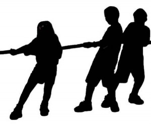 Work together like the muscles of the scapula Nothing cooperates like the muscles of the scapula. A single muscle acting alone, with even a small load, could distort this thin bone, causing it to crumple or become misaligned and the humerous to dislocate. To avoid this, the muscles always work together, simultaneously, holding the bone in a firm 360° embrace of muscular tension, keeping it in the correct alignment. Use the adult scapula to trace a rough outline of the bone on a large piece of cardboard. Use the vector diagram below, modified from Aiello and Dean (1990), to identify the key points of attachment for the cranking muscles; punch holes on the edges and tie on some 3-4 ft. strings. Now assign students to be the anchors (and activators) of the different muscles. Call out some movements. Rotate the scapula up and down. Don’t overpull or you’ll tear a muscle!
 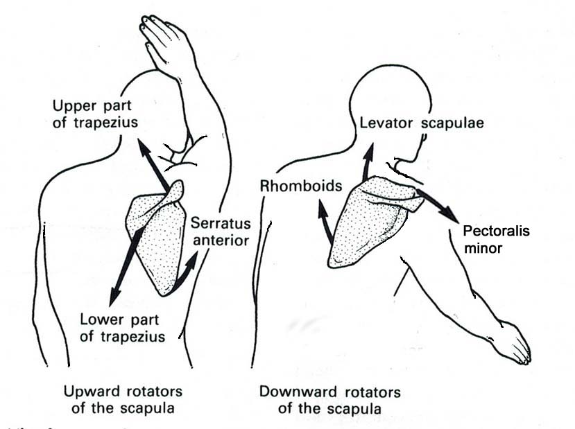 Hold the scapula level and balance a cup on top. Now try rotating. If anyone lets his or her line go slack the cup will fall. Can you do it without the cup falling (i.e. dislocating) like the head of the humerus? Your scapula muscles make these adjustments automatically with every move, in fact, often anticipating the arm movement. (Roberts, 1974) Talk about teamwork!
| Muscle | Action | Origin | Insertion |
| upper trapezius | upward rotator | cervical vertebrae | clavicle |
| lower trapezius | upward rotator | thoracic vertebrae | scapula spine |
| serratus anterior | upward rotator | ribs, front of chest | vertebral border |
| rhomboids | downward rotator | thoracic vertebrae | vertebral border |
| levator scapulae | downward rotator | cervical vertebrae | superior angle |
| pectoralis minor | downward rotator | ribs, front of chest | coracoid process |
Are you wondering what the other scapula muscles are doing? They originate on the scapula and serve to stabilize and control the head of the humerus or move the arm.
More things to do with the scapula
 Things to think about
Compare the scapulae. Are the juvenile scapulae simply miniature versions of the adult bone? It’s hard to tell. It may help to trace their outlines on paper using the adult bone as a model and draw in the missing edges. Scientists have suggested several different techniques for comparing scapulae but most start by measuring the length and width of the supraspinous and infraspinous fossae. The latter is more than twice as long in humans (Schultz, 1930), but here in Megalonyx (all three individuals, in fact) they are equal.* Remember, the fossae anchor the rotator cuff muscles; the power muscles attach on the edge where they can get better leverage. The only muscles inserting in the supraspinous region are the serratus anterior, which rotates the scapula up to help raise the arm (control is more important here since the arm isn’t that heavy) and the levitator scapulae, which rotates the scapula downward to boost the pulling power of the arm. Larger fossae and longer superior edges mean a more powerful arm, or an arm that is being used in a wider range of motion. (Roberts, 1974) If the supraspinous fossae of the”Baby” and “Toddler” are proportional to the adult’s, it’s a strong clue they were moving their arms like adults too. See more things to do with the scapula for more about calculating scapular fossae indices.
*Notably, Megalonyx shares that distinction with lions and just a few other carnivores. That probably aids in prey capture and possibly in their bounding running. (Roberts, 1974)
Scientists disagree about how much of the shape of the scapula is influenced by the functional demands placed on it. Some believe it is entirely shaped by the loads placed on it. (Preuschoft et al., 2010) Others point to research suggesting the bone is less plastic and is constrained by its phylogenetic heritage (i.e. ancestral genes). Monteiro and Abe (1999) calculated that almost half the shape variation was so caused in a sample of 17 species of Xenarthrans.
 Future research
If human experience is any indication juvenile sloths probably had to walk or crawl on all fours before they learned to stand upright and walk on two legs (if they did). Two sets of juvenile sloth scapulae at distinctly different stages of development may provide the clues for researchers to answer questions like, “When did toddler sloths learn to walk upright?”  “Did juvenile sloths walk on all fours or did they ride clinging to the backs of their mothers?” Other juvenile sloth scapulae have been found, but comparing them to adult scapulae is confounded by the fact they came from different geographical regions and eras, and the question of whether scientists were seeing genetic differences, functional differences or geographic variation. The Tarkio Valley sloth scapulae may hold the answers.
Conclusion
The scapula is about cranking the upper arm bone. Far from being a firm anchor for the humerus, scapulae rotate and contribute significantly both to the range of motion and the power of the upper limbs. The shape tells much about how an animal uses its limbs, and a committed quadruped is easily distinguished from a sometimes biped, and this scapula bespeaks unmatched power in every direction, both to pull and rotate. Most sloth scientists believe ground sloths evolved from arboreal ancestors (Monteiro and Abe, 1999), and with scapulae well-adapted for reaching while maintaining balance, and emphasizing power and flexibility over speed. They were pre-adapted for moving to the ground and standing upright. Many physical anthropologists believe humans followed the same evolutionary path. (Preuschoft, 2004) Did Megalonyx walk bipedally on the ground too or are the strong hints in the scapula merely the genetic legacy of its arboreal ancestors? We’ll have to look at other bones to know for sure.
TO LEARN MORE ABOUT OR TO BORROW THE UNIVERSITY OF IOWA MUSEUM OF NATURAL HISTORY GEO-2-GO DISCOVERY TRUNKS CALL OR CONTACT THE MUSEUM.
References
Aiello, L. and Dean, C. 1990. Introduction to Human Evolutionary Anatomy. Academic Press Limited. San Diego, CA.
Bass, W. M. (1971) Human Osteology: A laboratory and field manual of the human skeleton. University of Missouri. Columbia, MO.
Burke, R. M. 2008. Can we estimate stature from the scapula? A test considering sex and ancestry. Master’s Thesis, University of Idaho.
Campobasso, C. P., Di Vella, G. and Intona, F. Jr. 1998. Using scapular measurements in regression formulae for the estimation of stature. Journal of Biological Research 74: 75-82.
Churchill, S. E. and Trinkaus, E. 1990. Neandertal scapular glenoid morphoplogy. American Journal of Physical Anthropology 83: 147-160.
Ciochon, R. L. and Corruccini, R. S. 1977. The coraco-acromial ligament and projection index in man and other anthropoid primates. Journal of Anatomy 1254: 627-632.
Davis, D. D. 1949. The shoulder architecture of bears and other carnivores. Fieldiana, Zoology 31: 285-305.
Davis, D. D. 1964. The Giant Panda: A morphological study of evolutionary Mechanisms. Fieldiana, Zoology Memoirs 3: 1-364.
Educational Technology Clearinghouse, Florida Center for Instructional Technology, College of Education, University of Southern Florida http://etc.usf.edu/clipart/.
Fleagle, J. G. 1976. Locomotion and posture of the Malayan Siamang and implications for hominoid evolution. Folia Primatologica 26: 245-269.
Inman, V. T., Saunders, J. B. and L.C. Abbott. 1944. Observations on the functions of the shoulder joint. The Journal of Bone and Joint Surgery 26: 1-30.
McDonald, H. G. 1977. Description of the osteology of the extinct gravigrade edentate Megalonyx with observations on its ontogeny, phylogeny and functional anatomy. Master’s Thesis. University of Florida.
Monteiro, L. R. and Abe, A. S. 1999. Functional and historical determinants of shape in the scapulae of Xenarthran mammals: Evolution of a complex morphological structure. Journal of Morphology 241: 251-263.
Owen, R. 1842. Description of the Skeleton of an Extinct Giant Sloth, Mylodon robustus, Owen, with observations on the osteology, natural affinities, and probable habits of the Megatheroid quadrupeds in general. John Van Voorst, Publ. London.
Preuschoft, H. 2004. Mechanisms for the acquisition of habitual bepedality: are there biomechanical reasons for the acquisition of upright bipedal posture? Journal oif Anatomy 204: 363-384.
Preuschoft, H., Hohn, B., Scherf, H., Schmidt, M., Krause, C., Witzel, U. 2010. Functional analysis of thre primate shoulder. International Journal of Primatology 31: 301-320.
Roberts, D. 1974. Structure and function of the primate scapula. In Primate Locomotion, F. A. Jenkins, Jr. (ed.), Academic Press, NY. pp. 171-200.
Schultz, A. 1930. The skeleton of the trunk and limbs of higher primates. Human Biology 2: 303-438.
Smith, J. M. and R. J. G. Savage. 1956. Some locomotory adaptations in mammals. Journal of the Linnean Society of London, Zoology 42: 603-622.